Key details for this exam, checked against the published exam outline
Each question shows the correct answer and an explanation of why it is right
What is a normal response to dobutamine stress testing?
During dobutamine stress testing, a normal physiological response includes increased myocardial contractility leading to a decrease in left ventricular (LV) cavity size during systole due to more effective ejection. Concurrently, systolic blood pressure increases due to the inotropic and chronotropic effects of dobutamine.
An increase in LV cavity size during stress would suggest impaired contractility or ischemia, which is abnormal.
This normal response is detailed in the 'Textbook of Clinical Echocardiography, 6e', Chapter on Stress Echocardiography and Hemodynamic Responses20:400-405Textbook of Clinical Echocardiography.
Which pathology is consistent with the left ventricular strain pattern shown in this image?
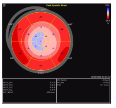
The strain imaging shown is a classic example of the 'apical sparing' pattern, highly characteristic of cardiac amyloidosis. In cardiac amyloidosis, the basal and mid segments of the left ventricle show markedly reduced longitudinal strain (represented here by more positive or less negative strain values), while the apical segments retain relatively preserved strain (more negative strain values). This 'cherry on top' or 'bull's eye' pattern with apical strain preserved distinguishes amyloidosis from other causes of LV dysfunction.
This pattern is not typical of apical hypertrophy, which would show focal thickening and abnormal strain limited to the apex. Non-ischemic cardiomyopathy generally has a more diffuse and uniform reduction in strain without the apical sparing. Right coronary artery infarcts affect the inferior and posterior walls and would have segmental strain abnormalities corresponding to the infarct distribution, not the typical apical sparing.
The left ventricular global longitudinal strain (GLS) in amyloidosis is typically severely reduced, but the relative preservation of apical strain is a hallmark useful for diagnosis, as described in the 'Textbook of Clinical Echocardiography, 6e' (Chapter on strain imaging and infiltrative cardiomyopathies) .
Which acute disease state is indicated with McConnell's sign?
McConnell's sign is an echocardiographic finding characterized by regional right ventricular (RV) dysfunction with akinesia of the mid-free wall but preserved contractility of the apex. This pattern is highly specific for acute pulmonary embolism (PE).
In acute PE, sudden obstruction of the pulmonary artery leads to acute right ventricular pressure overload, causing regional wall motion abnormalities. The sparing of the apex differentiates it from other causes of RV dysfunction such as myocardial infarction.
This sign is considered a useful bedside clue in the echocardiographic diagnosis of PE, especially when combined with clinical findings and Doppler evidence of elevated pulmonary pressures.
The sign is described in the 'Textbook of Clinical Echocardiography, 6e', Chapter on Acute Right Heart Dysfunction, with reference to McConnell's original description and its clinical significance in acute pulmonary embolism diagnosis20:340-345Textbook of Clinical Echocardiography.
Which of the following is commonly evaluated by the sniff maneuver?
Comprehensive and Detailed Explanation From Exact Extract:
The sniff maneuver is commonly used in echocardiography to assess right atrial pressure (RAP) indirectly by observing changes in the size and collapsibility of the inferior vena cava (IVC). During a sniff or rapid inspiration, negative intrathoracic pressure normally causes the IVC to collapse. The degree of IVC collapse during the sniff test correlates with RAP.
If the IVC is dilated and fails to collapse significantly with a sniff, this suggests elevated right atrial pressure, which can be caused by right heart failure, pulmonary hypertension, or volume overload.
This maneuver is not used to evaluate left atrial pressure or outflow tract obstructions, which require other echocardiographic parameters.
This assessment method is described in the 'Textbook of Clinical Echocardiography, 6e', Chapter on Right Heart Evaluation and Hemodynamics20:300-305Textbook of Clinical Echocardiography.
Which echocardiography assessment requires mitral inflow pulsed wave, pulmonary venous pulsed wave, and tissue Doppler of the mitral annulus?
Assessment of left ventricular diastolic function by echocardiography involves evaluating mitral inflow velocities with pulsed wave Doppler (E and A waves), pulmonary venous flow patterns (systolic and diastolic waves), and tissue Doppler imaging of the mitral annulus to measure early diastolic (e') velocities.
This combination allows differentiation of normal versus abnormal relaxation, elevated filling pressures, and grading of diastolic dysfunction. The myocardial performance index evaluates global ventricular function but does not specifically require these Doppler measures. Systolic function is assessed mainly by ejection fraction and wall motion. Mitral regurgitation severity uses color Doppler and vena contracta measurements.
This multiparameter diastolic function evaluation is outlined in the 'Textbook of Clinical Echocardiography, 6e', Chapter on Diastolic Function Assessment20:210-220Textbook of Clinical Echocardiography.
In cardiac tamponade, how do transvalvular pressure gradients change during expiration?
In cardiac tamponade, there is a characteristic reciprocal respiratory variation in transvalvular flow velocities due to ventricular interdependence and impaired cardiac filling. During expiration, the intrathoracic pressure increases, which leads to decreased right ventricular filling and thus decreased transtricuspid flow velocity. Simultaneously, left ventricular filling increases, causing an increase in transmitral flow velocity.
Therefore, during expiration, the transmitral gradient increases while the transtricuspid gradient decreases. This phenomenon reverses during inspiration, where transtricuspid flow increases and transmitral flow decreases. These respiratory variations are diagnostic hallmarks of tamponade physiology and help distinguish it from other conditions.
This principle is illustrated in Doppler echocardiographic studies of ventricular inflow and is described with diagrams in the 'Textbook of Clinical Echocardiography, 6e' (Chapter 10: Pericardial Disease), highlighting the changes in transmitral and transtricuspid velocities during the respiratory cycle in tamponade .
Exam domains verified against: Official ARDMS AE-Adult-Echocardiography exam guide, last checked October 2026.
Master normal cardiac structures, great vessels, anatomic variants, and the complete cardiac cycle from chambers to valves. You need to recognize normal hemodynamic responses to stress testing and document physiologic findings with Doppler interrogation, including normal valve gradients and arterial and venous waveforms.
Sample question from this domain above: Q3
This is nearly half the exam. You must know cardiac diseases including valve stenosis and regurgitation, cardiomyopathies, septal defects, arrhythmias, congenital anomalies like tetralogy of Fallot and Marfan syndrome, pericardial disease, and complications of myocardial infarction. Master postoperative findings for valve repairs, replacements, TAVR, and intracardiac devices.
Incorporate patient history and imaging data into your exam, position patients properly, apply EKG leads, manage blood pressure monitoring, and identify critical findings. Know contraindications for echocardiographic procedures and recognize medical emergencies including your role in managing them.
Perform measurements across 2D, 3D, M-mode, and Doppler including aortic valve planimetry, diastolic function parameters, left ventricular dimensions, and pressure half-time calculations. Execute standard views from apical, parasternal, subcostal, and suprasternal windows, and perform provocative maneuvers like Valsalva and handgrip.
Recognize and manage imaging artifacts in both 2D and Doppler, adjust console settings for optimal display and Doppler recording including harmonics, and apply ultrasound contrast agents safely. Understand the physical principles of contrast agents and their contraindications.
Sample question from this domain above: Q2
Common questions about the exam itself

