Key details for this exam, checked against the published exam outline
Each question shows the correct answer and an explanation of why it is right
Which vessel lies anterior to the uncinate process?
The superior mesenteric vein (SMV) lies directly anterior to the uncinate process of the pancreas. The uncinate process wraps around the posterior aspect of the SMV and SMA. The portal vein and IVC lie more posteriorly in relation to the pancreatic head.
According to Moore's Clinically Oriented Anatomy:
''The superior mesenteric vein crosses anterior to the uncinate process of the pancreas.''
Moore KL, Dalley AF, Agur AMR. Clinically Oriented Anatomy. 8th ed. Wolters Kluwer, 2018.
Gray's Anatomy for Students, 4th ed., Elsevier, 2019.
---
A patient presents with right lower quadrant pain and fever. Which condition is most likely indicated by the arrow on this image?
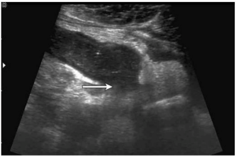
The ultrasound image demonstrates a tubular, non-compressible, blind-ending structure located in the right lower quadrant (RLQ) with associated echogenic periappendiceal fat and possibly adjacent fluid or phlegmon. These features are consistent with appendicitis. Given the clinical history of fever and RLQ pain, along with the irregular borders and complex periappendiceal findings, the diagnosis of a ruptured appendix is most likely.
Key sonographic features of ruptured appendicitis include:
Non-visualization or distortion of the normal appendiceal wall architecture
Periappendiceal fluid collection or abscess
Disruption of the echogenic submucosal layer
Surrounding fat stranding (hyperechoic inflammatory changes)
Clinical correlation with fever and peritonitis
Comparison of answer choices:
A . Bowel obstruction typically shows dilated bowel loops with air-fluid levels, not a tubular structure like the appendix.
B . Intussusception presents with a target or ''donut'' sign in a transverse view, not a linear tubular structure.
C . Enlarged lymph nodes are usually round or oval and hypoechoic with a central echogenic hilum, without a tubular appearance.
D . Ruptured appendix --- Correct. The ultrasound features and clinical presentation match.
Rumack CM, Wilson SR, Charboneau JW, Levine D. Diagnostic Ultrasound, 5th ed. Elsevier; 2017.
Jeffrey RB, Laing FC, Townsend RR. Acute appendicitis: sonographic criteria based on 250 cases. Radiology. 1988;167(2):327--329.
American Institute of Ultrasound in Medicine (AIUM) Practice Parameter for the Performance of the Ultrasound Examination for Appendicitis (2020).
Which sonographic finding is commonly associated with transitional cell cancer of urinary bladder?
Transitional cell carcinoma (TCC) typically presents as a non-mobile, polypoidal, focal intraluminal mass projecting from the bladder wall. Mobility of the lesion helps differentiate TCC from blood clots or debris.
According to Rumack's Diagnostic Ultrasound:
''Bladder TCC most often appears as a non-mobile, polypoid mass attached to the bladder wall.''
Rumack CM, Wilson SR, Charboneau JW, Levine D. Diagnostic Ultrasound. 5th ed. Elsevier, 2017.
AIUM Practice Parameter for Bladder Ultrasound, 2020.
---
Which organ is held in place by the lienorenal, gastrosplenic, and phrenocolic ligaments?
The spleen is suspended in the left upper quadrant by several peritoneal ligaments, including:
Lienorenal (splenorenal) ligament --- attaches spleen to the left kidney.
Gastrosplenic ligament --- attaches spleen to the stomach.
Phrenocolic ligament --- supports the spleen inferiorly between diaphragm and colon.
These ligaments stabilize the spleen's position while allowing some mobility.
According to Moore's Clinically Oriented Anatomy:
''The spleen is connected to the stomach by the gastrosplenic ligament and to the posterior abdominal wall (near the left kidney) by the splenorenal (lienorenal) ligament. The phrenocolic ligament provides inferior support.''
Moore KL, Dalley AF, Agur AMR. Clinically Oriented Anatomy. 8th ed. Wolters Kluwer, 2018.
Gray's Anatomy for Students, 4th ed., Elsevier, 2019.
---
A lactating female presents with a tender, swollen breast, erythema, and fever. Which condition is most likely present in this image?
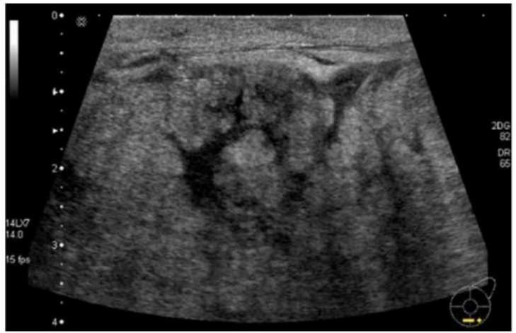
The clinical presentation---tender, swollen breast with erythema and fever---in a lactating female strongly suggests acute mastitis. The sonographic findings support this diagnosis. In the image, the breast parenchyma shows diffuse, hypoechoic, and heterogeneous echotexture with increased vascularity, which is consistent with inflammatory changes typical of mastitis.
Mastitis is a common complication during lactation, particularly in the first few weeks postpartum. It results from milk stasis and subsequent bacterial infection, commonly due to Staphylococcus aureus. Ultrasound features of mastitis include:
Ill-defined, hypoechoic, edematous areas in the breast parenchyma
Increased Doppler flow due to hyperemia
Skin thickening
Ductal dilatation may also be present
If left untreated, mastitis may progress to abscess formation, which would appear as a localized, complex fluid collection with peripheral hyperemia and internal debris. However, the image does not show a well-formed fluid collection consistent with abscess.
Option B (Ductal carcinoma): Inappropriate here due to the acute clinical scenario and patient age. Ductal carcinoma typically presents as a hypoechoic mass with irregular margins and posterior shadowing, not diffuse edema or inflammatory changes.
Option D (Galactocele): This benign milk-filled retention cyst typically appears anechoic or with fluid--fluid levels but lacks signs of inflammation and systemic symptoms such as fever.
Option A (Abscess): This could be a differential, but abscesses usually present with a well-defined anechoic or complex mass. The absence of a discrete collection and the diffuse appearance makes mastitis more likely.
Mendelson EB. Practical Ultrasound: An Illustrated Guide. Springer, 2004. Chapter: Breast Ultrasound.
American College of Radiology (ACR). ACR Practice Parameter for the Performance of a Breast Ultrasound Examination, 2022.
Rumack CM, Wilson SR, Charboneau JW, Levine D. Diagnostic Ultrasound. 5th Edition. Elsevier, 2018. Chapter: Breast, pp. 1169--1175.
Which condition is most consistent with the findings in the image below?
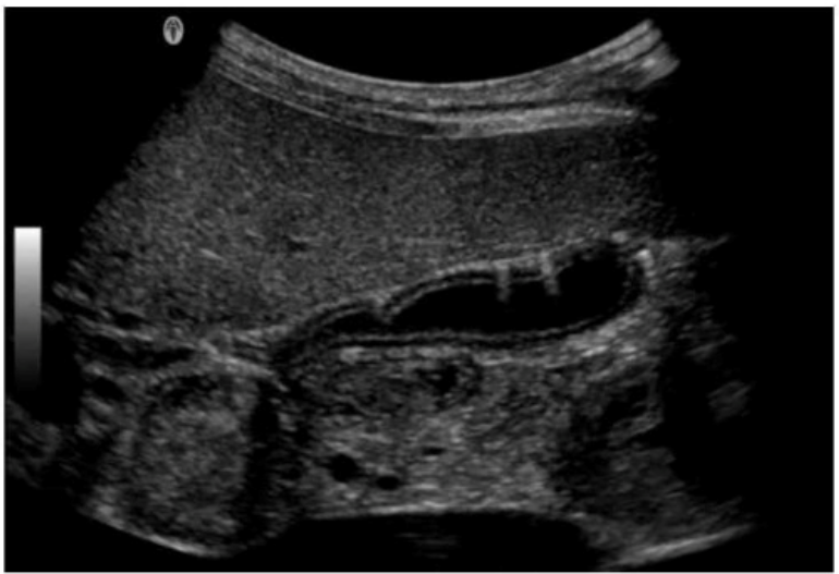
The ultrasound image shows echogenic foci with dirty shadowing and reverberation artifacts within the gallbladder wall and lumen. These features are characteristic of emphysematous cholecystitis, a severe, life-threatening variant of acute cholecystitis caused by gas-forming organisms (e.g., Clostridium or E. coli) infecting the gallbladder wall.
Sonographic features of emphysematous cholecystitis:
Echogenic gas within the gallbladder wall or lumen
Reverberation or 'dirty' shadowing artifacts
May show intramural gas bubbles or 'ring-down' artifact
Often seen in diabetic or immunocompromised patients
No gallstones may be present ('acalculous cholecystitis')
Clinical context:
More common in elderly men and diabetics
Presents with right upper quadrant pain, fever, and leukocytosis
Surgical emergency due to risk of perforation and sepsis
Differentiation from other options:
A . Adenomyomatosis: Involves gallbladder wall thickening with ''comet tail'' artifacts due to Rokitansky-Aschoff sinuses, not intramural gas.
B . Porcelain gallbladder: Shows curvilinear calcification of the gallbladder wall --- dense echogenic rim with posterior shadowing.
C . Gangrenous cholecystitis: May show wall irregularity, intraluminal membranes, and absence of Doppler flow but lacks intramural gas.
Rumack CM, Wilson SR, Charboneau JW, Levine D. Diagnostic Ultrasound. 5th Edition. Elsevier, 2018. Chapter: Gallbladder and Biliary System, pp. 155--160.
American College of Radiology (ACR). Appropriateness Criteria for Right Upper Quadrant Pain, 2022.
Radiopaedia.org. Emphysematous cholecystitis: https://radiopaedia.org/articles/emphysematous-cholecystitis
Exam domains verified against: Official ARDMS AB-Abdomen exam guide, last checked October 2026.
Assess the physical characteristics of anatomic abdominal structures including organs, blood vessels, and soft tissues. Evaluate perfusion patterns and organ function using sonographic imaging.
Identify and characterize pathologic processes in abdominal structures. Recognize vascular abnormalities and assess for trauma-related changes and complications following surgical procedures.
Apply concepts of ultrasound equipment function and image optimization to produce diagnostic quality images. Apply knowledge of imaging artifacts and their sources to improve diagnostic accuracy.
Incorporate clinical data and patient history into sonographic assessment and reporting. Obtain accurate measurements and maintain quality standards while assisting with or supporting diagnostic and procedural studies.
Sample question from this domain above: Q5
Common questions about the exam itself

